
Vasa Praevia: The Hidden Challenge in Pregnancy
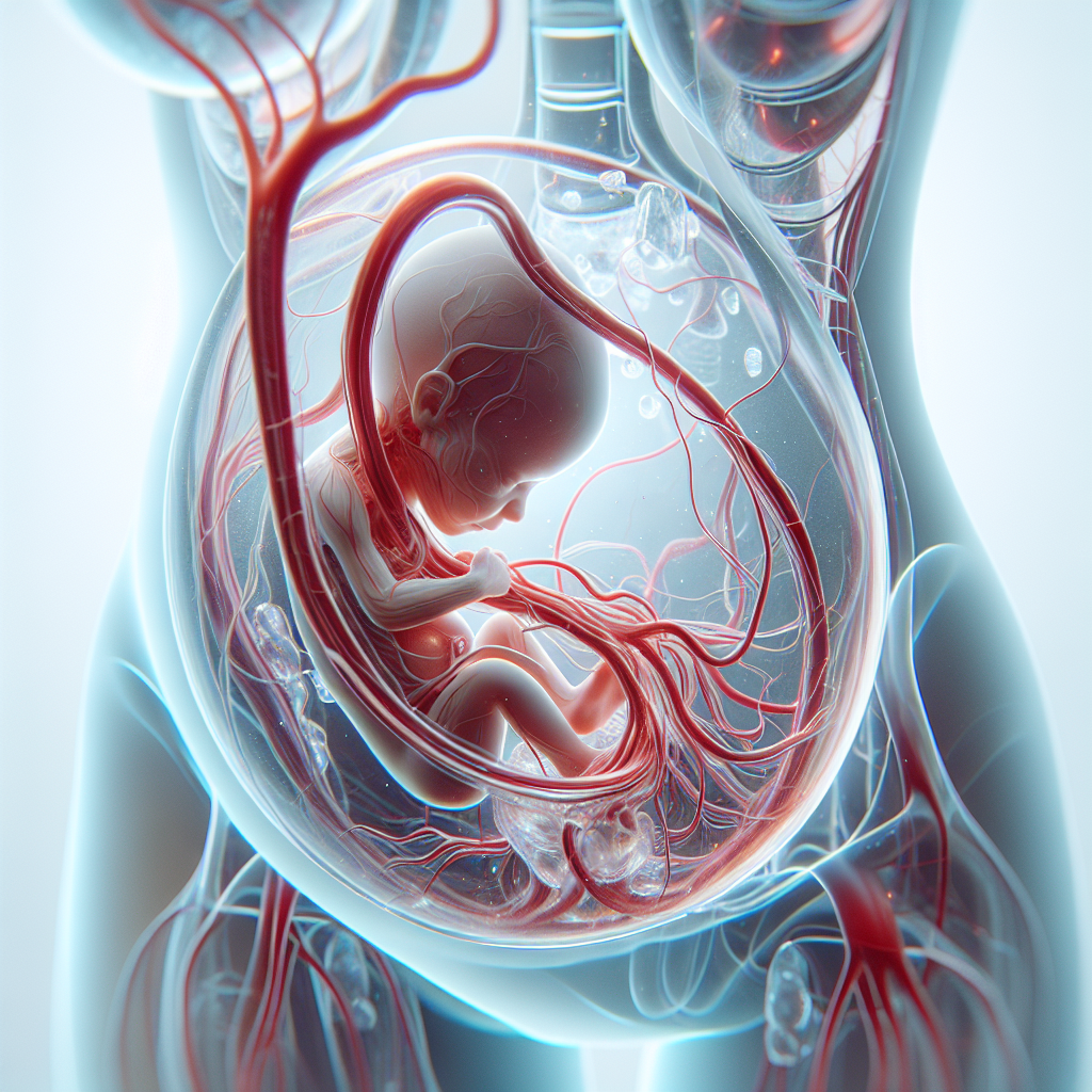
Imagine a hidden challenge during pregnancy that can be as thrilling as a mystery novel, yet as serious as a medical emergency. Vasa praevia is a rare but critical condition that occurs when fetal blood vessels cross or run near the internal opening of the uterus, beneath the baby. This condition can be a significant concern during childbirth, as these vessels are at risk of rupturing when the membranes break, leading to potentially life-threatening bleeding for the baby. Vasa praevia is typically diagnosed during the second or third trimester of pregnancy through ultrasound, and it requires careful monitoring and planning for delivery to ensure the safety of both mother and child.
The "who" in this scenario includes expectant mothers and their healthcare providers, who must be vigilant in identifying and managing this condition. The "what" is the presence of fetal blood vessels in a precarious position, which can be detected through advanced imaging techniques. The "when" is during pregnancy, particularly in the later stages, when the risk of complications increases. The "where" is within the uterus, specifically at the cervix, where these vessels can be compressed or torn during labor. The "why" is the critical need to prevent fetal blood loss, which can occur rapidly and with severe consequences if not addressed promptly.
Vasa praevia is a condition that underscores the importance of prenatal care and the advancements in medical imaging that allow for early detection. With proper diagnosis and a well-coordinated delivery plan, the risks associated with vasa praevia can be significantly reduced, ensuring a safe outcome for both mother and baby. This condition highlights the incredible journey of pregnancy and the intricate dance of biology and medicine that supports it.